










How we Gruve
Our solutions are designed for speed to scale
AI Platform
Private AI infrastructure, production-ready in 2 weeks. Sovereign AI stack with zero data egress, GPU orchestration, and 30% cost advantage.

WHY GRUVE
Built for enterprise at scale



Video
AI-Powered Security Operations Center (SOC)
Gruve's AI-powered Security Operations Center combines AI agents with human security experts to deliver faster, more accurate protection. AI agents triage alerts, investigate routine incidents, and hunt threats around the clock, while experienced analysts focus on judgment calls that need a human touch.
Success stories
Make data-driven decisions





Industries we serve in India

Manufacturing

Manufacturers, industrial operators, and supply chain networks often operate across many plants, systems, and legacy equipment. AI transformation assessments help teams discover where automation and AI can improve operations, monitor process gaps, and prioritize initiatives that affect production and business continuity.
- Discover legacy systems, manual processes, and automation opportunities.
- Monitor operational gaps across plants and supply chain systems.
- Give leaders clearer reporting for prioritization and planning.

Government

Government agencies, public sector bodies, and municipal services often operate across many legacy systems, departments, and citizen-facing platforms. AI transformation assessments help teams discover where AI can improve service delivery, identify outdated processes, and prioritize initiatives that affect public trust and operational continuity
- Discover legacy systems, manual workflows, and automation opportunities.
- Identify data silos and gaps in cross-department visibility.
- Give leaders clearer reporting for prioritization and budget planning.

Telecom

Telecom operators, network providers, and communications companies often operate across many networks, systems, and customer touchpoints. AI transformation assessments help teams discover where AI can improve operations, monitor
network and process gaps, and prioritize initiatives that affect service reliability and business continuity.
- Discover legacy systems, manual processes, and automation opportunities.
- Monitor operational gaps across network and customer-facing systems.
- Give leaders clearer reporting for prioritization and planning.

BFSI

Banks, capital markets firms, payment providers, and insurers often operate across many platforms, products, and regulatory environments. AI transformation assessments help teams discover where automation and AI can add value,
monitor operational gaps, and prioritize initiatives that affect customer trust and business continuity.
- Discover manual processes, fragmented data, and automation opportunities.
- Monitor operational gaps and third-party AI dependencies.
- Give leaders clearer reporting for prioritization and compliance.

Healthcare

Hospitals, clinics, healthtech platforms, pharma, and insurance providers often operate across many public-facing systems and patient touchpoints. AI transformation assessments help teams discover where AI can improve caredelivery, monitor
operational gaps, and prioritize initiatives that affect patient outcomes and business continuity.
- Discover manual workflows, fragmented data, and automation opportunities.
- Monitor operational gaps across clinical and administrative systems.
- Give leaders clearer reporting for prioritization and compliance.

Technology

Software companies, SaaS platforms, and technology providers often operate across many products, teams, and customer-facing systems. AI transformation assessments help teams discover where AI can improve products and operations,
monitor technical gaps, and prioritize initiatives that affect customer experience and business continuity.
- Discover fragmented tooling, manual processes, and automation opportunities.
- Monitor technical debt and gaps in product AI readiness.
- Give leaders clearer reporting for prioritization and roadmap planning.
Solutions to AI challenges
-
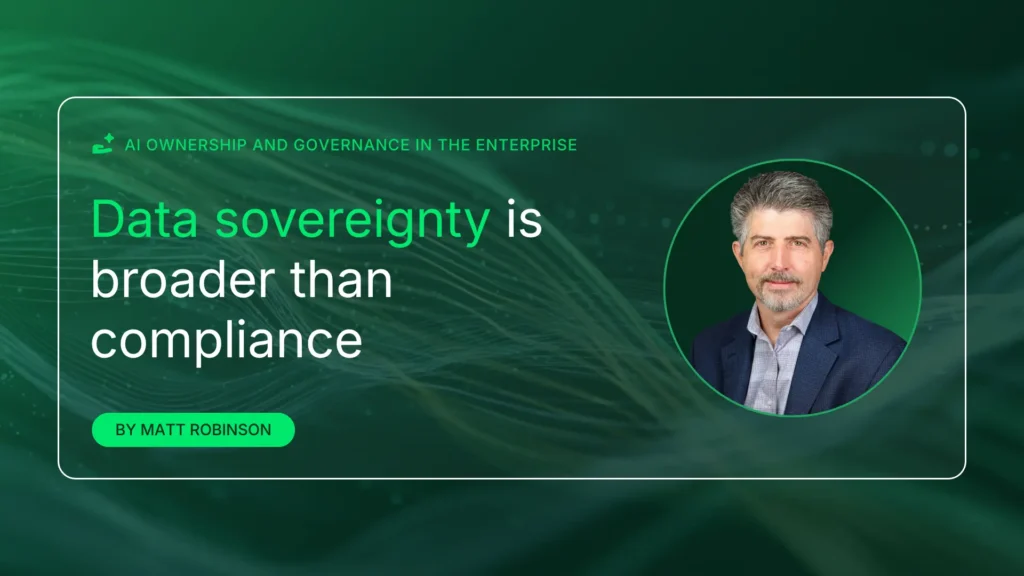

Data sovereignty is broader than compliance
Data sovereignty usually gets pulled into the AI compliance conversation, which is understandable. If the data includes payment information, protected health information,…
-
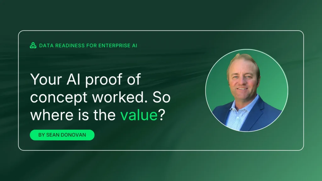

Your AI proof of concept worked. So where is the value?
The pilot-to-production wall is usually a data problem—not an AI problem. Before I joined Gruve, I worked with a company on an…
-


Making enterprise AI operational
There is a practical question I keep hearing from customers and partners. A customer completes a successful AI pilot. As the organization…
-


Cloud incident response services
What earlier took 285 minutes now takes just 72 minutes: We are talking about data exfiltration. In 2026, AI has reimagined and…
Unlock your true speed to scale
Enable scalable, secure, and measurable AI execution in production. Start with a discovery call.